DNA to Protein
PlaygroundThe others with no size
A gene is a stretch of DNA that spells out a protein.
- Shelf
- Tiny World
- Kind
- Built from a recipe
- Your own
- Your own DNA
- Sound
- Yes
- Licenses
- Public domain, CC0
How to play, in the Playground
Tap to make the protein. Pick a gene, add a mutation, or type your own DNA.
Its controls
| Control | Kind | Range | Start |
|---|---|---|---|
| Make the protein | Tap | ||
| Make the protein | Button | ||
| Gene | List | 5 choices | Hemoglobin beta (HBB) |
| Mutation | List | 4 choices | None |
| At codon | Setting | ||
| Base in the codon | Setting | ||
| New base | List | 4 choices | T |
About it
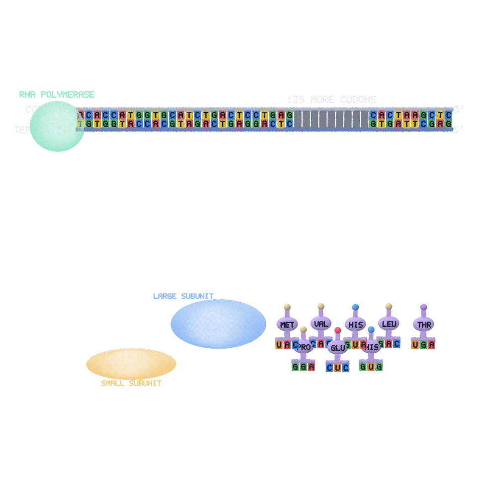
A gene is a stretch of DNA that spells out a protein. Tap and RNA polymerase opens the double helix and reads the template strand, building messenger RNA that matches the coding strand (with U for T). A ribosome then reads the mRNA three bases (a codon) at a time from the start codon AUG; each tRNA whose anticodon pairs with the codon brings its amino acid, and the chain grows until a stop codon. The chain then folds into the protein's real shape from the Protein Data Bank.
The genes are real (NCBI); only the start and end are drawn base by base. Try a mutation: changing base 2 of codon 7 in hemoglobin beta to T is the sickle-cell change (glutamic acid to valine).
Details
- Splats
- Opens at 200,000 at Mid Detail, 59,991 at Low, and up to 200,000 at Max
- Your own files
- .fa, .fasta, .fna, .txt, .seq
Is it right?
The splash shows a real gene being transcribed into mRNA and translated into a protein, which then folds into its measured structure. The bases, codons, amino acids and structures are real data, and the genetic code is the standard one, checked by tests. The machines (RNA polymerase, the ribosome, tRNAs) are drawn as simple shapes, much smaller and much faster than the real ones, and most of each gene is shortened.
Correct
The genetic code is the standard code: 64 codons, 61 for amino acids and 3 stop codons (UAA, UAG, UGA); AUG codes for methionine and starts the protein.1
The code table is NCBI's translation table 1 written in its usual order; translation starts at the first ATG and reads in threes to the first stop codon in the same frame.
Correct
Each sample gene's coding sequence translates exactly into its protein: hemoglobin beta (147 amino acids), preproinsulin (110), lysozyme (148) and green fluorescent protein (238).2345
tools/tw2-genes.mjs takes the CDS from each NCBI record and stops unless the splash's own translation equals the record's /translation; a test checks it again.
Correct
RNA polymerase reads the template strand; the mRNA is built 5′ to 3′, complementary to the template, so it matches the coding strand with U in place of T.6
The coding strand is drawn on top (5′ at the left), the template under it (3′ at the left), and each mRNA base appears under its template base as the complementary RNA base.
Simplified
The ribosome's small subunit with the initiator tRNA (methionine) binds near the mRNA's 5′ cap and scans to the first AUG; the large subunit then joins.7
The small subunit and the first tRNA come to the 5′ end together, move along the leader to the start codon, and the large subunit comes down onto them.
Simplified
Each tRNA's anticodon pairs with its codon in the A site; the chain moves onto it (a peptide bond), the ribosome moves one codon, and the empty tRNA leaves from the E site.7
A tRNA carrying the codon's amino acid docks with its anticodon on the codon, the chain shifts from the P-site tRNA to it, the ribosome moves three bases along, and the tRNA two places back leaves.
Correct
At a stop codon no tRNA binds; a release factor frees the finished chain.7
When the stop codon reaches the A site, a tRNA-shaped release factor comes in, the chain leaves the tunnel, and the subunits come apart.
Simplified
The finished chain folds into the protein's measured structure (hemoglobin beta from PDB 4HHB, insulin from 1MSO, lysozyme from 1LZ1, GFP from 1GFL).891011
Each amino acid moves to the place of its alpha carbon in the PDB entry, matched to the protein by sequence (GFP by residue number); amino acids missing from a crystal (GFP's first residue and last eight) hang loosely from their neighbors.
Simplified
The cell cuts pieces away: hemoglobin beta's first methionine, insulin's signal peptide (1-24) and its C-peptide with the basic pairs around it (55-89), and lysozyme's signal peptide (1-18).121314
These amino acids drift away as one piece while the rest folds.
Correct
Changing base 2 of codon 7 of HBB from A to T turns GAG (glutamic acid) into GTG (valine): the sickle-cell change, Glu6Val in the finished protein's numbering.15
The mutation is made in the sequence before transcription, so it shows in the DNA, the mRNA, the codon, the tRNA and the amino acid; the panel line gives both numbers.
Correct
Adding or removing one base shifts the reading frame: every codon after it changes, and translation usually stops early at a new stop codon.16
The splash reads the changed sequence (on into the real 3′ untranslated region if it must) to the first stop in the new frame; a chain cut short like this is not folded.
- 1The Genetic Codes: 1. The Standard Code, NCBI Taxonomy
- 2Homo sapiens hemoglobin subunit beta (HBB), mRNA, NM_000518.5, NCBI Nucleotide
- 3Homo sapiens insulin (INS), transcript variant 1, mRNA, NM_000207.3, NCBI Nucleotide
- 4Homo sapiens lysozyme (LYZ), mRNA, NM_000239.3, NCBI Nucleotide
- 5Aequorea victoria green-fluorescent protein mRNA, complete cds, M62653.1, NCBI Nucleotide (Prasher and others, Gene 111:229-233, 1992)
- 6Biology 2e, 15.2 Prokaryotic Transcription, OpenStax
- 7Biology 2e, 15.5 Ribosomes and Protein Synthesis, OpenStax
- 84HHB: the crystal structure of human deoxyhaemoglobin at 1.74 Å resolution, RCSB Protein Data Bank
- 91MSO: T6 human insulin at 1.0 Å resolution, RCSB Protein Data Bank
- 101LZ1: refinement of human lysozyme at 1.5 Å resolution, RCSB Protein Data Bank
- 111GFL: structure of green fluorescent protein, RCSB Protein Data Bank
- 12P68871 (HBB_HUMAN), UniProt
- 13P01308 (INS_HUMAN), UniProt
- 14P61626 (LYSC_HUMAN), UniProt
- 15HBB gene, MedlinePlus Genetics (U.S. National Library of Medicine)
- 16Biology 2e, 15.1 The Genetic Code, OpenStax
- Simplified
- Only the start and end of each gene are drawn base by base: five bases before the start codon, the first seven codons, the last codon and the stop codon. The codons in between are drawn as plain bases and go by quickly, and a label says how many there are.
- Simplified
- The genes are drawn as coding sequences (like a cDNA). The real human genes have introns, which are spliced out of the RNA in the nucleus before it leaves for the cytoplasm; the splash leaves out splicing, the poly(A) tail and the trip through the nuclear pore, and most of the 5′ leader.
- Simplified
- DNA is drawn as a flat ladder; the real double helix turns once every 10.5 base pairs. The transcription bubble is drawn a few bases wide; in the cell it is about a dozen.
- Simplified
- RNA polymerase and the ribosome are drawn far smaller than they are next to the DNA and mRNA (a real ribosome covers about 30 bases of mRNA), as clear shells so you can see inside. The exit tunnel is drawn holding about five amino acids; a real one holds 30 or more.
- Simplified
- The new chain gathers in a loose ball over the ribosome and folds after its release. In the cell, many proteins begin to fold while they are being made, often helped by chaperones, and hemoglobin and insulin finish as parts of larger assemblies (with heme, zinc and partner chains).
- Simplified
- The story takes about half a minute. A cell makes a protein of 150 amino acids in well under a minute, and the transcription and translation of one gene happen many times over, by many polymerases and ribosomes at once.
Sources and credits
| Work | By | License |
|---|---|---|
| NCBI RefSeq NM_000518.5 (HBB), NM_000207.3 (INS), NM_000239.3 (LYZ) and GenBank M62653.1 (GFP) | the National Center for Biotechnology Information | Public domain |
| RCSB Protein Data Bank entries 4HHB, 1MSO, 1LZ1 and 1GFL (alpha carbons) | the wwPDB and the structures' authors | CC0 1.0 |